I would appreciate that!
Sal
joined 3 years ago
MODERATOR OF
Thanks for the explanation. I will look into how to remove cellular contents and how to separate layers. I didn't know this could be achieved with acid :D
Cool, thanks!
In this picture the cells in the epidermis layers appear to lack chloroplasts. I wonder if that's the case... That might also explain this.

Very cool. Before staining, did you prepare it in some way?
Great album cover!!
Is this with a filter? A stain? Post-processing?
I think so... What is interesting to me though is that the chloroplasts (I think) are visible in the stomata. Do you think it might be because the layers below it are thinner? Actually... What is "under" the stomata that opening up allows air to access to? Rhetorical question - I am just thinking out loud. I'll look it up!
Ok, I have looked at this leaf diagram but I still don't get why the chloroplasts in guard cells would be more visible if the layers below are so chlorophyll-dense.... Need to go deeper....

Thanks!
I didn't know about microcystins. Interesting molecules! I think this particular species does not produce them, but I won't eat them... at least for now. I might grow some edible algae in the future.
That's not the cyanobacteria's fault, I think that it is mine, so thanks for pointing that out. In the post-processing I clicked on the microscope slide to white balance because the background seemed too blue. But now that you mention this I think that the bluish hue in the background was realistic and setting it to white removed the blue from the blue-green cyan. Without the white balance the color is more cyan:

Dendrologist?
I didn't even need to tear the leaf, I just placed a drop of oil on top and used the 100x objective. The green is from the chlorophyll, yes. I suspect that the cells are so packed with chloroplasts that it just looks smoothly green.
11
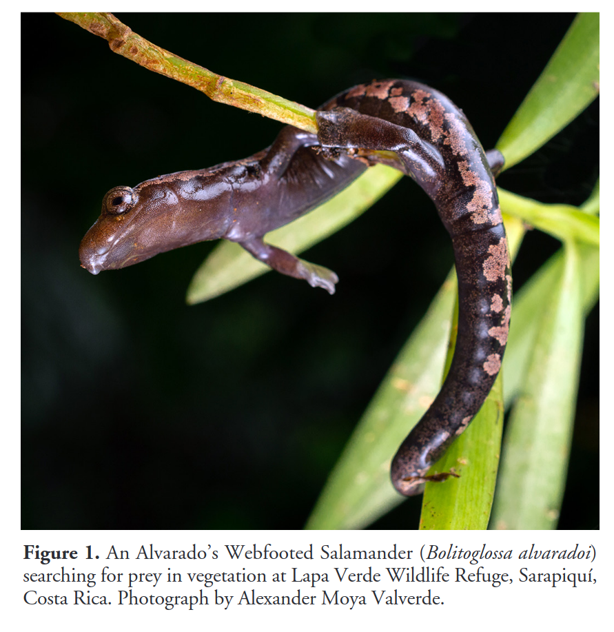
[PDF] Alvarado’s Webfooted Salamander, Bolitoglossa alvaradoi: Activity, hunting behavior, and prey selection in Costa Rica
(journals.ku.edu)
A new paper on a Bolitoglossid showed up in my alerts today! 🥳
It is a one-pager and comes with a beautiful photo of a foraging arboreal salamander 😄
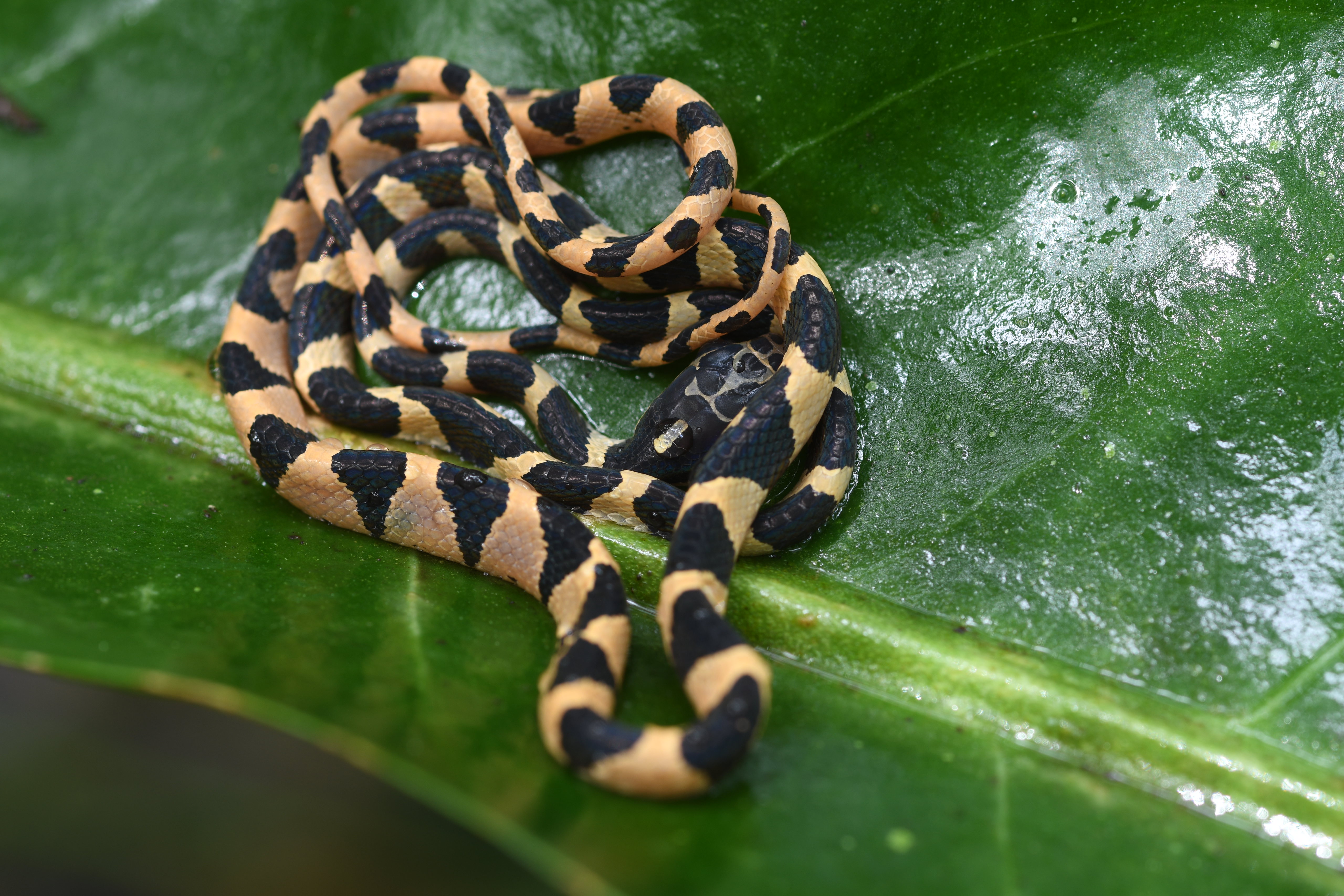
Found this Imantodes cenchoa sleeping on top of a leaf in Punta Laguna, Quintana Roo.
Here is a photo of where it was sleeping:

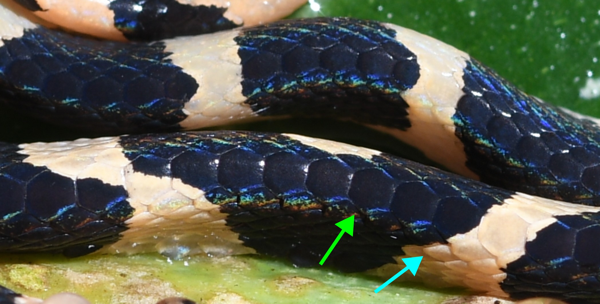
There are a few different snakes of the genus Imantodes in Yucatán. What sets this species apart is that the row of scales on its back consists of enlarged scales. Here is a closeup emphasizing that row of scales. The green arrow points at an enlarged mid-dorsal scale, the blue arrow at a regular scale.
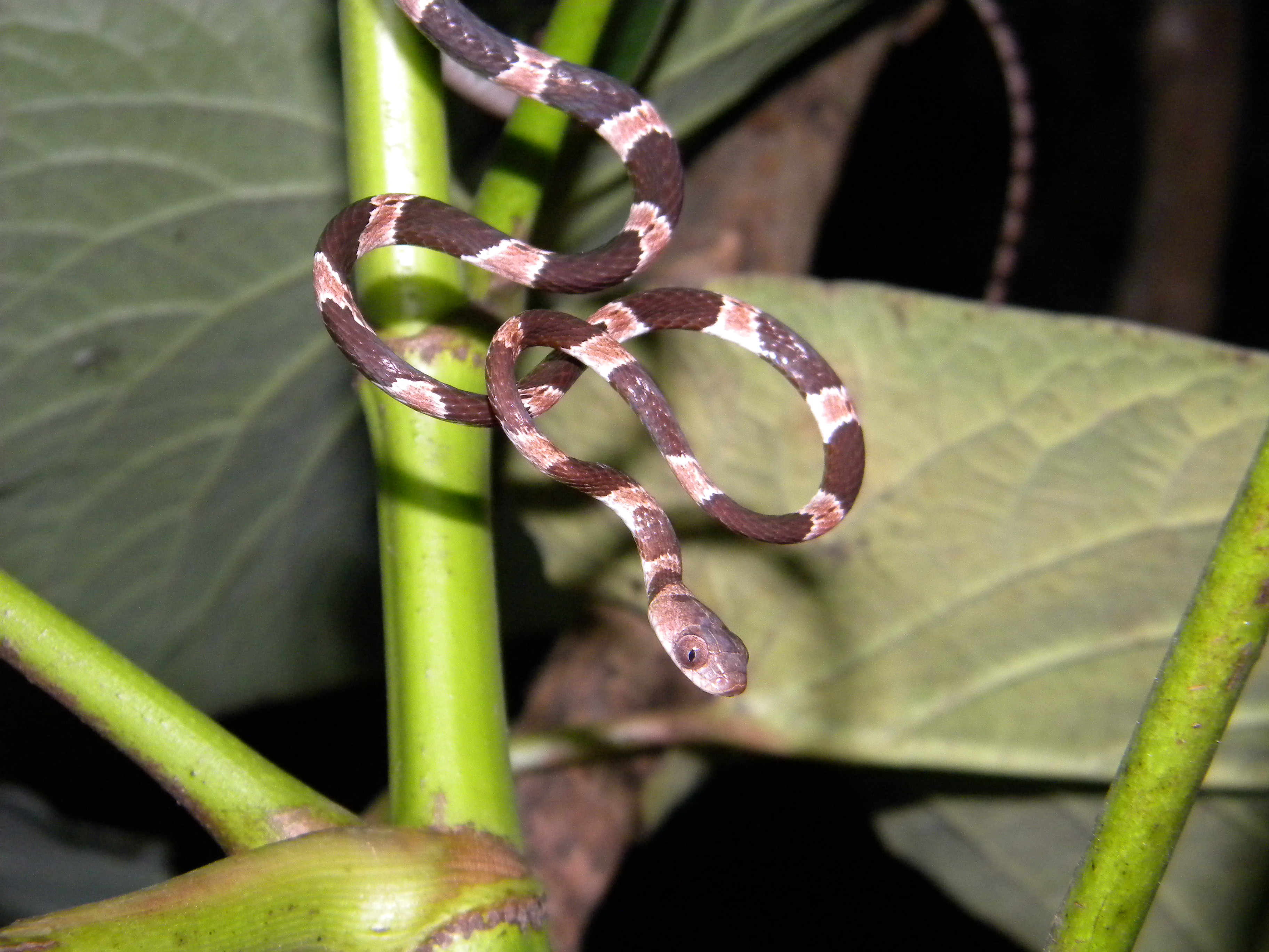
In comparison, here is a photo from an Imantodes tenuissimus that I took back in 2009 in Mérida, Yucatán (with a less sharp camera), and a closeup of its mid-dorsal scales that does not show this enlargement.

Permanently magnetized